Which Conditions Can Be Confused with Vitiligo?
Which Conditions Can Be Confused with Vitiligo?

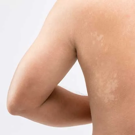
Vitiligo is an autoimmune skin condition characterized by the loss of melanin pigment, resulting in distinct white patches on the skin. However, not all white patches indicate vitiligo. Several dermatological conditions may present with similar depigmented or hypopigmented lesions, especially in their early stages. For this reason, an accurate diagnosis requires a detailed differential diagnostic approach by a dermatologist.
Visual similarity to vitiligo does not necessarily mean identical underlying mechanisms. While vitiligo involves immune-mediated destruction of melanocytes, other conditions may stem from infections, inflammation, or genetic factors. Misdiagnosis can lead to ineffective or even harmful treatments, delayed intervention, and increased emotional stress for the patient.
1. Pityriasis Alba
Commonly seen in children and adolescents, pityriasis alba presents as light-colored, mildly scaly patches, typically on the face. Key distinguishing features include:
- Partial pigment loss, unlike the total depigmentation of vitiligo
- Mild or no itching, sometimes with slight flaking
- Associated with atopic dermatitis history
- Often self-resolving without treatment
While easily confused with early vitiligo, pityriasis alba is benign and non-progressive.
2. Tinea Versicolor
A superficial fungal infection caused by Malassezia furfur, it appears as white, pink, or brownish spots, often with fine scaling. Typical features:
- Affects chest, shoulders, back, and neck
- Triggered by heat, humidity, and sweating
- Lesions may coalesce and are usually asymmetrical
- Responsive to antifungal treatment
Unlike vitiligo, pigmentation typically returns after fungal control.
3. Postinflammatory Hypopigmentation
This occurs after inflammatory skin conditions like eczema, psoriasis, or trauma (e.g., burns, laser treatments). Features include:
- History of inflammation, rash, or skin injury
- Incomplete loss of melanin; melanocytes remain intact
- Pigment loss is irregular and reversible over time
It can mimic vitiligo but is usually temporary and improves spontaneously.
4. Nevus Depigmentosus
A congenital, non-progressive white patch present from birth. It is:
- Stable in size over time
- Not associated with inflammation or immune activity
- Shows mild fluorescence under Wood’s lamp (unlike vitiligo’s bright white)
- Often isolated and not familial
It is a harmless pigment anomaly and not related to vitiligo.
5. Halo Nevus
An immune reaction causes a white ring (halo) to form around a mole (nevus). This condition:
- Typically arises during puberty
- May lead to the disappearance of the mole itself
- Can occur alone or in association with vitiligo
- Requires observation if multiple halos are present
Though it shares immune mechanisms, halo nevus has a distinct clinical behavior.
6. Idiopathic Guttate Hypomelanosis
Seen in middle-aged or older adults, especially on the shins, arms, and forearms, this condition presents as:
- Small (less than 5 mm), drop-like white macules
- Associated with sun exposure and aging
- No itching or pain
- Non-progressive and cosmetic in concern only
It differs from vitiligo by being static and much smaller in size.
7. Albinism
A congenital, genetic disorder where melanin production is globally impaired, affecting skin, hair, and eyes. Unlike vitiligo:
- Present since birth
- Involves widespread depigmentation
- Associated with eye abnormalities like photophobia or nystagmus
- Caused by enzyme deficiency, not immune destruction
Albinism is clearly distinct in presentation and cause.
Key Differences Between Vitiligo and Similar Conditions
|
Feature |
Vitiligo |
Mimickers |
|
Onset |
Childhood to early adulthood |
Varies: birth, childhood, or late age |
|
Lesion color |
Milky white, sharply defined |
Pale, scaly, or diffuse |
|
Progression |
Often spreads |
Usually stable or self-limiting |
|
Wood’s lamp test |
Bright white fluorescence |
Weak or no fluorescence |
|
Symptoms |
Usually asymptomatic |
May include scaling or itching |
Frequently Asked Questions
Fungal conditions like tinea versicolor cause flaky patches that respond to antifungal treatments, whereas vitiligo does not involve fungus and does not improve with antifungals.
No. Conditions like nevus depigmentosus are present from birth and do not progress, unlike vitiligo.
A Wood’s lamp examination, detailed dermatologic assessment, and in some cases, a skin biopsy confirm the diagnosis.
No. Several conditions can resemble vitiligo. Proper medical evaluation is essential for correct diagnosis.
No. However, vitiligo patients may have an increased risk for other autoimmune conditions, so regular checkups are advisable.