Could Skin Whitening Be a Sign of Vitiligo?
Could Skin Whitening Be a Sign of Vitiligo?

Does Skin Whitening Always Mean Vitiligo?
Skin discoloration or light patches often raise concerns about vitiligo, but not all whitening is caused by this autoimmune condition. Vitiligo is characterized by the complete loss of pigment (depigmentation) due to the destruction of melanocytes. However, many other conditions can cause similar-looking light patches or hypopigmentation.
Proper evaluation of the color, shape, duration, and distribution of the lesion is crucial to determine whether it’s vitiligo or another skin condition.
Common Causes of Skin Whitening
Skin lightening can result from either hypopigmentation (reduced pigment) or depigmentation (complete pigment loss). Vitiligo is a classic example of depigmentation, but here are other common causes:
1. Pityriasis Alba
- Common in children
- Pale, slightly scaly patches, usually on the face
- Often follows mild inflammation
- Tends to resolve spontaneously
2. Tinea Versicolor (Fungal Infection)
- Caused by Malassezia yeast
- Patches appear white, pink, or brown
- May be mildly itchy
- Treated with antifungal medications
3. Post-inflammatory Hypopigmentation
- Follows eczema, psoriasis, acne, or skin injuries
- Temporary and fades over time
- No treatment typically required
4. Chemical Leukoderma
- Results from contact with certain toxic chemicals
- Irregular, asymmetrical patches
- Usually in areas of repeated exposure
5. Albinism
- A genetic condition affecting the entire body
- Complete absence of melanin in skin, eyes, and hair
- Diagnosed at birth
Recognizing these differences helps distinguish them from early vitiligo.
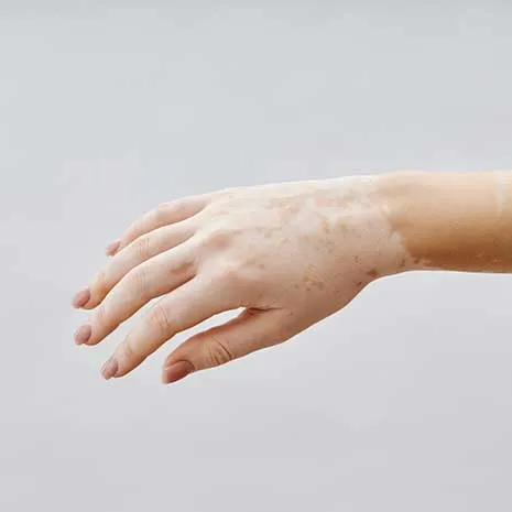
Early Signs of Vitiligo
Vitiligo often begins as small, milky-white patches with sharply defined borders. In early stages, lesions may go unnoticed—especially in fair-skinned individuals—until sun exposure makes them more visible.
Signs Suggestive of Vitiligo:
- Well-demarcated white patches
- Loss of hair pigment in the area (leukotrichia)
- Lesions at sites of trauma (Koebner phenomenon)
- Family history of vitiligo or autoimmune diseases
- Symmetrical distribution (e.g., both hands or both feet)
- Bright white fluorescence under Wood’s lamp
Presence of these features increases the likelihood of a vitiligo diagnosis.
The Role of Wood’s Lamp in Diagnosis
A Wood’s lamp emits UVA light and is used in dark rooms to enhance visibility of pigment loss. It’s a key tool for detecting early or subtle vitiligo lesions.
In Vitiligo:
- Lesions glow bright white or bluish
- Boundaries appear more distinct
- Helpful in differentiating from fungal or post-inflammatory hypopigmentation
When Should You See a Dermatologist?
Consult a dermatologist if you notice:
- A white patch lasting longer than two weeks
- Lesions that are spreading or changing
- Family history of vitiligo or other autoimmune disorders
- Hair whitening within the patch
- History of trauma or chemical exposure in the area
- Lesions with clear, symmetrical borders
Early diagnosis allows for better monitoring and timely treatment if necessary.
Differentiating Vitiligo from Other Causes
|
Condition |
Lesion Appearance |
Itchiness |
Spontaneous Resolution |
|
Vitiligo |
Sharp, milky-white |
No |
Rare |
|
Pityriasis Alba |
Pale, slightly scaly |
Mild |
Yes |
|
Tinea Versicolor |
Light or dark patches |
Mild |
Yes (with treatment) |
|
Chemical Leukoderma |
Irregular white patches |
No |
No |
|
Post-inflammatory |
Follows a prior rash or injury |
No |
Yes |
Frequently Asked Questions
It could be, but many other conditions cause similar symptoms. A dermatological exam is necessary for accurate diagnosis.
No. Several non-vitiligo conditions—especially in children and adolescents—can cause temporary skin whitening.
No. Vitiligo lesions are typically non-itchy and painless.
Yes. Some causes, like pityriasis alba or post-inflammatory hypopigmentation, are reversible.
A Wood’s lamp examination is the most common first-line tool. In uncertain cases, skin biopsy and blood tests may be used.